
 mariusmoza
Otitis Cytology in Dogs: A Vet’s Guide to Ear Cytology Interpretation
Otitis externa is one of the most common reasons dogs and cats end up in the exam room, and it rarely resolves with a single visit. A VetCompass study of dogs under UK primary veterinary care found a one-year period prevalence of 7.30% across a random sample of more than 22,000 dogs, with breed and ear carriage ranking as the highest risk factors. Feline cases occur at a lower but still clinically meaningful rate.
Otitis cytology in dogs is what turns that exam room presentation into a treatment decision. Getting the diagnosis right the first time depends on it, but staining variability and turnaround time can slow the workflow down at exactly the moment a clear answer matters most.
This post breaks down how to interpret ear cytology findings, how to tell Malassezia from cocci on the slide, what is actually driving diagnostic inconsistency between clinics, and how faster, more standardized slide prep is changing that workflow for practices that see these cases every week.
Why Cytology Is the Standard for Otitis Externa Diagnosis
Otitis externa is a multifactorial disease. Effective, lasting treatment depends on separating primary causes (like allergies or parasites) from secondary infections and the perpetuating factors that keep an ear from healing even after the underlying cause is addressed, a framework outlined in the Journal of the American Veterinary Medical Association’s review on managing recurrent otitis externa.
Bacterial culture is sometimes used to guide therapy, but it has real limitations in the ear. A 2022 study published in Veterinary Sciences compared cytology, bacterial culture, and 16S amplicon profiling in dogs with chronic suppurative otitis externa and found only moderate agreement between cytology and culture results, around 60% of samples. The same study noted that over a third of the bacterial species detected through DNA sequencing at meaningful abundance levels were missed by culture entirely. Culture and susceptibility panels are also built around blood-level drug concentrations, which do not reflect the much higher concentrations achieved with topical therapy, one reason many dermatologists rely on cytology as the primary diagnostic and monitoring tool rather than defaulting to culture.
Cytology, by contrast, gives an immediate read on what is actually present on the slide: cocci, rods, yeast, and the inflammatory response driving clinical signs. That immediacy is exactly why the Merck Veterinary Manual lists cytological evaluation, alongside history and otoscopic exam, as a core part of otitis externa diagnosis, and why it should be performed at the initial visit and at every recheck, not just when a case looks complicated. Accurate ear cytology interpretation at each of those visits is what actually drives the treatment decision, not the culture result that may take days to come back.
Ear Cytology Interpretation: What You Are Looking For on the Slide
A useful ear cytology read typically covers a few key findings:
What you see | What it suggests | Clinical takeaway |
Cocci | Often Staphylococcus species | Usually more straightforward to resolve with topical therapy |
Rods | Commonly Pseudomonas or other gram-negatives | Signals a more complicated case; may need a different treatment approach and closer monitoring |
Yeast (Malassezia) | Frequent contributor in both dogs and cats | Shapes topical therapy choice; often present alongside bacteria, so a slide is not fully read until both are accounted for |
Neutrophils (especially degenerated) | Active bacterial process | Higher counts support more aggressive treatment |
Few to no inflammatory cells | Case may be resolving, or not truly infectious | Worth considering allergic otitis rather than infection |
Intracellular organisms | More established, significant infection | May warrant more aggressive treatment than extracellular organisms alone |
Organism density (few vs. sheets) | Distinguishes early/resolving cases from active infection | Density, not just presence, helps determine whether topical therapy is still needed |
When rods dominate the slide, the stakes go up since these organisms are often associated with greater antimicrobial resistance concerns. A retrospective analysis of bacterial isolates from dogs with pyoderma and otitis externa found that while Staphylococcus species, particularly S. pseudintermedius, were the most common isolates, Pseudomonas and Proteus were also frequently identified, and multidrug resistance was a recurring finding. The authors recommend prioritizing topical therapy and reserving systemic antibiotics for exceptional cases after susceptibility testing, which is another argument for cytology as the tool that guides the topical plan.
What Counts as an Otitis Externa Cytology Positive
This is where practices diverge more than most clinicians realize. There is no single agreed threshold for calling an ear cytology positive. Commonly cited cutoffs for yeast and bacteria per high-power field vary between references and between individual dermatologists, and none of them are universally adopted.
That means the call often comes down to the reader’s judgment of density, morphology, and the inflammatory picture, applied to whatever the slide happens to look like. Which makes slide quality the variable that quietly decides borderline cases.
None of this is useful if the sample itself is inconsistent. A comparative study of two ear cytology collection methods in dogs with otitis externa found that a cotton-tipped swab technique and a deep-canal aspiration technique produced different yields of neutrophils, a reminder that consistent sampling matters just as much as consistent interpretation. A well-collected sample that then sits on an inconsistently stained slide still leaves room for two clinicians to read the same ear differently. found that a cotton-tipped swab technique and a deep-canal aspiration technique produced different yields of bacteria, yeast, and inflammatory cells, a reminder that consistent sampling matters just as much as consistent interpretation. A well-collected sample that then sits on an inconsistently stained slide still leaves room for two clinicians to read the same ear differently.
Produced statistically different yields of neutrophils, a reminder that
Malassezia vs. Cocci on Ear Cytology: Telling Them Apart and Why It Changes Treatment
The single most common read in ear cytology is distinguishing yeast from bacteria, and most slides contain both.
Malassezia are unmistakable once you have seen a few. They are roughly 3 to 8 micrometers, several times the size of a coccus, and shaped like a peanut, a snowman, or a bowling pin depending on which textbook you learned from. That shape comes from broad-based unipolar budding, where the daughter cell separates from one pole without pinching off into a neat sphere. They take up Romanowsky stain readily and appear deep blue to purple. The ACVD clinical consensus guidelines on the biology, diagnosis, and treatment of Malassezia dermatitis in dogs and cats are the references worth keeping on hand for anything beyond identification.
Cocci are round, roughly one micrometer, and typically appear in pairs, tetrads, or grape-like clusters. At 100x oil they read as fine purple dots against the background, and the size difference next to a Malassezia organism is dramatic enough that the two are rarely confused once both are on the same slide.
The trouble is what mimics them. Melanin granules, keratin debris, and stain precipitate all produce small dark specks that can pass for cocci to a tired eye at the end of a long day. Stain precipitate in particular tends to show up when reagents are aging or dip times drift, which means the artifact most likely to be misread as bacteria is generated by the same staining inconsistency described in the next section.
Why the distinction drives treatment: yeast and bacteria require different active ingredients in the topical. A slide read as yeast-only leads to an antifungal-weighted product, a bacteria-only read leads to an antibacterial one, and the mixed infections that make up a large share of otitis cases need both addressed. Treating only the organism that jumped out first is one of the more common reasons an ear improves partially and then stalls.
Why Ear Cytology Results Vary Between Techs
Even with good sampling, staining and slide prep introduce variability that has nothing to do with a clinician’s skill. Manual Romanowsky staining involves several timed dip steps, and small differences in dip time, reagent freshness, or how a slide is dried can all change how the same sample looks under the microscope. Two technicians staining the same sample, on the same day, can produce slides that look meaningfully different, which can shift how a borderline case gets read and treated, especially when organism counts are close to a threshold that determines whether a case is called infected or not.
That inconsistency is a systemic issue built into manual staining workflows, not a reflection of individual technique or effort. It shows up most in high-volume practices, where the same tech may be staining dozens of slides a day, and in multi-doctor practices, where different clinicians may be reading slides prepared by different team members. It is also one of the harder problems to catch, since an inconsistent slide does not announce itself. It just quietly nudges a borderline read one way or the other, and the vet interpreting it usually has no way to know which one they are looking at.
This is the gap that standardized, automated slide processing is designed to close.
In-House Ear Cytology vs. Send-Out: What You Gain and What You Give Up
Most practices are performing ear cytology in-house, but here are some factors to consider.
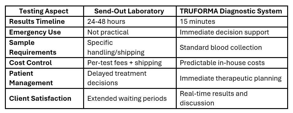
Send-out gets you a board-certified clinical pathologist reading the slide, which is genuinely valuable for ambiguous cases, suspected neoplasia, or anything that does not fit the usual otitis picture. What it costs you is time. The traditional workflow runs prepare, package, ship, wait, review, receive report, and the ear cytology turnaround time on that sequence is typically measured in days. For a patient who is painful and an owner who wants an answer before they leave, that gap is the whole problem.
In-house ear cytology collapses that timeline to the length of the appointment. The vet sees the slide, makes the call, and dispenses the medication topical before the client walks out. What you give up is the specialist read and, in a manual staining workflow, the consistency that a dedicated lab brings to slide prep.
For most otitis cases the clinical question is straightforward enough that in-house wins on speed without giving up much accuracy. The cases where send-out earns its turnaround are the ones that do not look like routine otitis in the first place. The practical answer for a lot of practices is not choosing between them, but having a workflow where the routine cases resolve in-house and the unusual ones escalate to a pathologist without restarting the sample collection process.

How the TRUVIEW® AI Microscope Speeds Up Ear Cytology Confirmation
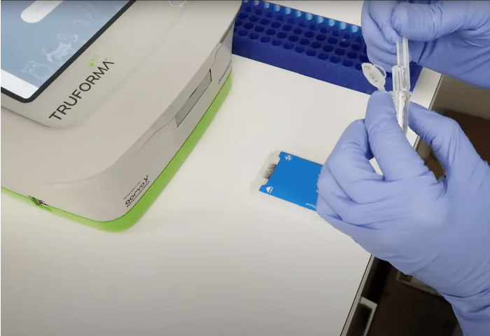
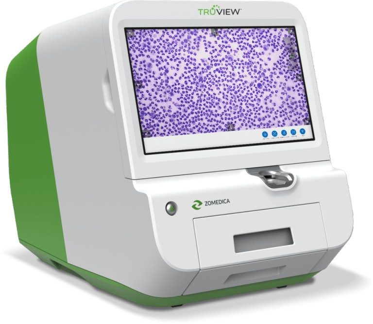
The TRUVIEW® AI microscope replaces the manual, timed-dip staining process with TRUprep® automated slide preparation. Insert the slide, and the system smears, stains, washes, dries, and digitizes it on a fixed cycle, so stain exposure, reagent volume, and drying are identical on every slide regardless of who is running the sample or how busy the clinic is that day. Instead of a tech eyeballing dip counts between appointments, every slide goes through the same standardized steps, which removes the tech-to-tech and day-to-day variability described above from the read itself.
For ear cases specifically, the Ear Cytology Quick Scan protocol streamlines slide prep and scanning so a slide that might otherwise sit in a queue behind other cases is imaged and ready to read in minutes. For a practice running multiple otitis cases in a single day, that difference in speed adds up: same-visit answers instead of asking an owner to wait for a callback, and less time between sample collection and a treatment decision.
The TRUVIEW system also captures and stores a digital image of every slide, which solves a second problem manual cytology does not: there is no permanent record of what the tech or vet actually saw under the scope at the time. A stored image can be pulled back up at a recheck, compared side by side with the current slide, or shared with a referring vet or a board-certified pathologist through the myZomedica® portal without needing to physically send a slide. That is a meaningful upgrade over a written note that says “moderate cocci, few WBCs” with no way to verify it later.
That telepathology connection is also what makes the in-house vs. send-out tradeoff less of a tradeoff. Routine cases resolve during the appointment, and the unusual ones go to a pathologist from the same digitized slide, without recollecting a sample or shipping anything.
To be clear on scope: the TRUVIEW microscope supports and speeds up cytology-based confirmation. The veterinarian is still the one interpreting the slide and making the diagnosis. The platform’s role is removing the staining variability and turnaround-time bottleneck from that process, not replacing clinical judgment.
Ready to see how the TRUVIEW microscope’s ear cytology workflow fits into your practice? Request a demo!
Chronic Ear Infection in Dogs: Why Cytology Consistency Matters Most Here
Chronic otitis is where repeatable cytology matters most. Rechecks are common in these cases, since resolving the underlying cause often takes multiple rounds of treatment adjustment, and each recheck depends on an accurate read to know whether the current plan is working.
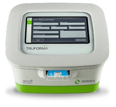
Working up a chronic ear infection in a dog means cytology is only part of the picture. Allergic disease is the most common primary cause, and the 2023 AAHA Management of Allergic Skin Diseases in Dogs and Cats Guidelines lay out the workup for allergic otitis externa alongside the rest of the minimum dermatologic database. Endocrine disease is another recurring culprit, and the TRUFORMA® diagnostic platform runs the thyroid and adrenal assays that help diagnose at the point of care.
This is also where inconsistent staining does the most damage. If a slide from the first visit was read as heavily infected and a recheck slide two weeks later reads as only mildly infected, is that real improvement or a difference in how the two slides were stained? Without standardization, that question is hard to answer with confidence, and it is exactly the kind of ambiguity that can lead to a treatment plan being extended, changed, or stopped based on noise rather than signal.
A fast, standardized cytology read at each recheck makes it easier to track whether an ear is actually improving or whether the treatment plan needs to change, without adding another 24-hour turnaround to a case that is already dragging on for the owner and the patient.
Frequently Asked Questions About Ear Cytology in Dogs
How many yeast per HPF is abnormal on ear cytology?
There is no universally agreed threshold, which is part of why slide-to-slide consistency matters so much. Commonly cited references suggest that more than 5 to 10 Malassezia organisms per high-power field (HPF) at 400x is considered significant, but individual dermatologists vary, and clinical signs should always factor into the interpretation. A high yeast count in an asymptomatic ear reads differently than the same count in a dog that has been scratching for weeks.
What does a normal ear cytology look like?
A cytology from a healthy ear typically shows low numbers of Malassezia (fewer than 5 per HPF), no bacteria, and little to no inflammatory response. Small amounts of ceruminous debris and occasional epithelial cells are expected. The absence of cocci, rods, and neutrophils is what distinguishes a normal slide from an early or resolving infection.
How often should you recheck ear cytology?
For active infections, a recheck cytology at two to three weeks is standard practice to confirm the infection is responding to treatment. For chronic or recurrent cases, cytology at every recheck visit is warranted regardless of how the ear looks clinically, since a slide that appears improved visually does not always match what is happening at the cellular level. Persistent organisms or a shift in organism type (cocci to rods, for example) between visits is clinically significant information that a visual exam alone will miss.
The Bottom Line
A quick recap for the slide in front of you:
- Rods, high organism density, or intracellular bacteria mean a more complicated case. Treat and monitor closely.
- No improvement between visits means questioning whether it is the treatment or the read before extending or switching therapy.
- Yeast and bacteria together means both need to be addressed in the treatment plan, not just the more obvious one.
- Sparse findings with ongoing signs means considering allergic otitis rather than infection.
Otitis is one of the most common problems in small animal practice. While it is often a one-visit diagnosis, it is rarely a condition that requires just one vet visit. Getting cytology interpretation right, consistently and quickly, is not just a diagnostic nicety. It is central to getting these patients comfortable faster, giving owners a clear answer sooner, and keeping cases from cycling back through the exam room unresolved.
© 2026 Zomedica Inc. All rights reserved. TRUVIEW is a registered trademark of Zomedica Inc.
 ecallen